Figure 1.
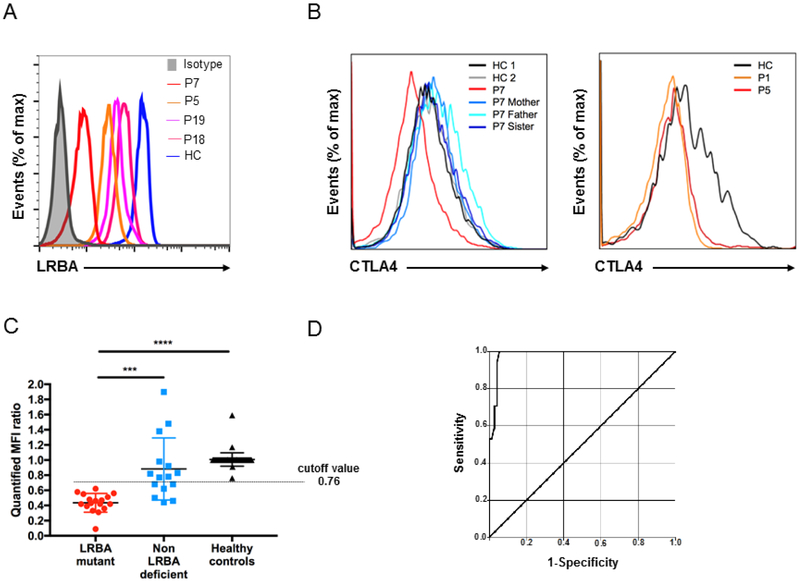
LRBA-deficient patients have low or absent LRBA and CTLA4 protein expression. A, LRBA expression in lymphocytes from LRBA-deficient patients, P7 (red line), P5 (orange line), P19 (purple line) and P18 (pink line) compared with a healthy control (blue line). B, Flow cytometric analysis demonstrates low CTLA4 expression on CD4+FOXP3+ Treg cells in LRBA-deficient patients P1, P5, and P7 compared to healthy controls (HC) and unaffected family members. C, LRBA mutant patients (red dots) have significantly decreased mean fluorescence intensity (MFI) ratio of LRBA expression in peripheral blood mononuclear cells compared to non-LRBA-deficient (blue dots) and healthy controls (black dots). D, Receiver operating characteristic (ROC) curve analysis shows the sensitivity and specificity of flow cytometric analysis for the detection of LRBA-deficient patients. The analysis was conducted on 17 mutation-verified LRBA-deficient patients, 15 patients who presented with a LRBA phenotype but had no LRBA mutation and 57 healthy controls. Area under curve was yielded as 0.98 with a 95% CI (0.96-1.00). *** p<.001 and **** p<.0001, Student unpaired 2-tailed t-test.

