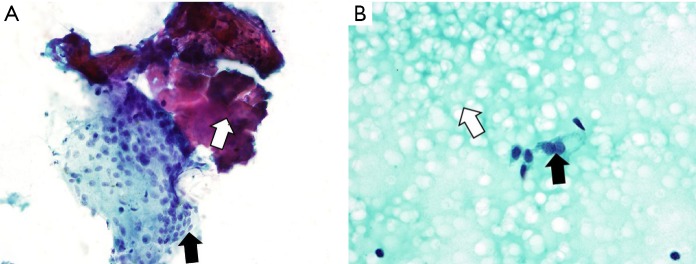
Figure 1.
Illustration of fine needle aspiration biopsy findings of a colloid nodule showing (A) (Papanicolaou stain, ×100) a fragment of inspissated colloid (white arrow) associated with a multinucleated foreign body giant cell (black arrow) and (B) (Papanicolaou stain, ×100) abundant thin watery colloid (white arrow) with few benign thyroid follicular cells (black arrow).

