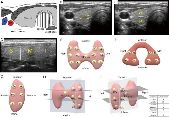
Figure 1.
Ultrasonographic views of thyroid gland and different localization methods of PTC tumors. (A) Diagram of thyroid anatomy; (B) in the longitudinal view, the gland was divided into superior (S), middle (M), and inferior (I) positions; (C) in the coronal view, the gland was divided into lateral (L) and central (C) positions; (D) in the sagitta view, the gland was divided into anterior (A) and posterior (P) positions; (E) longitudinal coronal location. The gland was divided into superior central (SC), superior lateral (SL), middle central (MC), middle lateral (ML), inferior central (IC), inferior lateral (IL) and isthums positions; (F) longitudinal sagittal location. The gland was divided into superior anterior (SA), superior posterior (SP), middle anterior (MA), middle posterior (MP), inferior anterior (IA), inferior posterior (IP) and isthmus positions. (G) Sagittal coronal location. The gland was divided into anterior central (AC), anterior lateral (AL), posterior central (PC), posterior lateral (PL) and isthmus positions; (H) 3D location in anteroposterior view. The superior anterior central (SAC), superior anterior lateral (SAL), middle anterior central (MAC), middle anterior lateral (MAL), inferior anterior central (IAC), inferior anterior lateral (IAL) and isthmus positions were shown; (I) 3D location in posteroanterior view. The superior posterior central (SPC), superior posterior lateral (SPL), middle posterior central (MPC), middle posterior lateral (MPL), inferior posterior central (IPC), inferior posterior lateral (IPL) and isthmus positions were shown. IJV, internal jugular vein; CA, carotid artery.

