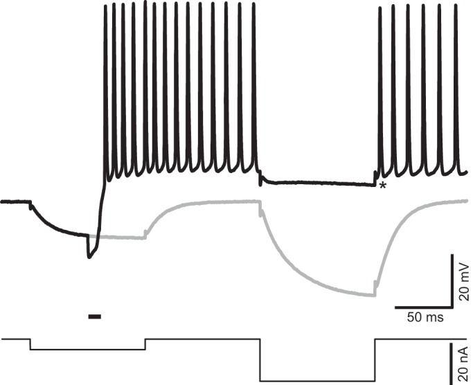
Fig. 5.

Ultrasound (US)-induced depolarization is associated with a reduction in membrane resistance. US-induced responses were investigated with a double current step protocol (bottom) including two 100-ms current steps −5 and −20 nA in amplitude, respectively, separated by an interval of 100 ms. The 2 current steps generated passive membrane responses in the absence of US stimulation (gray). A US burst delivered during a first current step induced repetitive firing (black). This firing was silenced by the second hyperpolarizing step, which only generated a small hyperpolarization. Asterisk identifies the baseline used to measure the amplitude of hyperpolarization (−4 mV) that silenced the firing. Horizontal bar above the current trace indicates the timing of US stimulation, namely, 10 ms at 2.0 MHz, and intensity of spatial peak temporal average = 7.7 mW/cm2.
