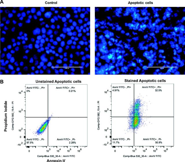
Fig. 6.
Hoechst 33342 staining of normal and apoptotic cells. A: control cells show normal nuclear staining, while the cells undergoing apoptosis demonstrated apoptotic chromatin changes: blebbing, fragmentation, and condensation under a fluorescence microscope at ×20. Apoptotic cells showed typical morphological features as DNA condensation, fragmentation, and nuclear shrinkage in UV light-exposed cells. B: flow cytometric analysis of annexin V-FITC/PI double-staining: Apoptotic cells collected after UV light treatment were incubated with Annexin V-FITC and/or propidium iodide (PI) and analyzed by flow cytometry. Annexin V+/PI− cells are early apoptotic cells; Annexin V+/PI+ cells are late apoptotic.

