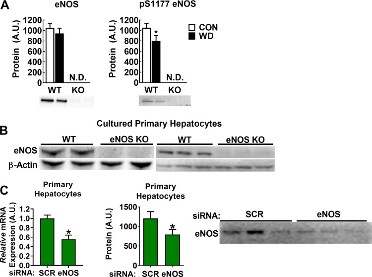
Fig. 1.
Endothelial NO synthase (eNOS) in liver tissue and primary hepatocytes. A: representative Western blot for eNOS and phosphorylated (p-)S1177 eNOS in liver homogenates from wild-type (WT) and eNOS knockout (KO) mice fed either control (CON) or Western diet (WD), n = 7–8/group. B: Western blot in cultured primary hepatocyte lysates from WT and eNOS KO mice. Blots on the left and right are from independent experiments (n = 5/genotype). C: small interfering (si)RNA-mediated knockdown of eNOS mRNA (left) and protein content (right) in WT hepatocytes; n = 7/group of independent replicates. A.U., arbitrary units; N.D., not detected; SCR, scramble. *P < 0.05 by paired two-tailed t-test.

