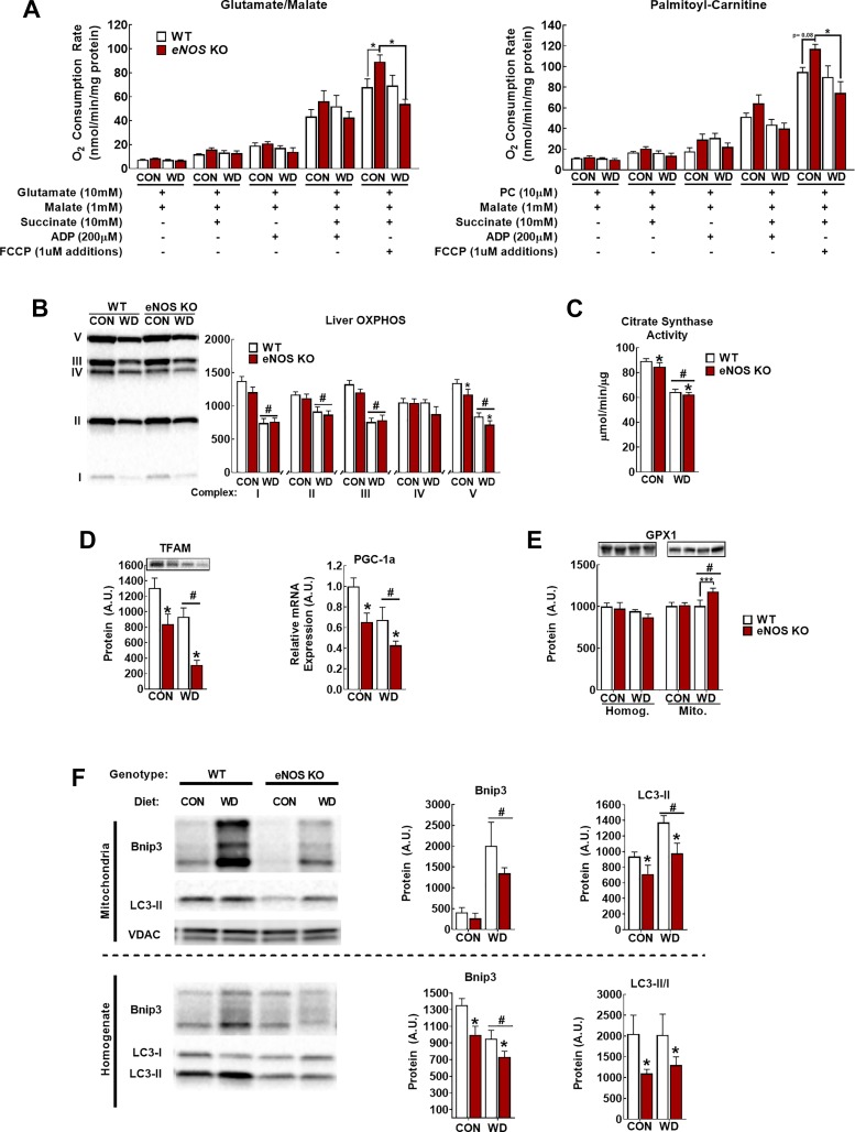
Fig. 4.
Endothelial NO synthase knockout (eNOS KO) alters mitochondrial function and turnover. A: glutamate/malate- and palmitoylcarnitine (PC)-supported respiration. B–C: assessment of mitochondrial content by oxidative phosphorylation (OXPHOS; B) and citrate synthase activity (C). D: markers of mitochondrial biogenesis peroxisome proliferator-activated receptor-γ coactivator 1α (PGC-1α) mRNA and mitochondrial transcription factor A (TFAM) protein. E: total liver (left) and mitochondrial (right) glutathione peroxidase-1 (GPX-1) content. F: representative Western blot images and densitometric quantification of BCL-2-interacting protein-3 (BNIP3) and light-chain 3B (LC3-II) in the mitochondrial fraction (top) and whole liver homogenate (bottom). VDAC, voltage-dependent anion-selective channel protein. Note that the arrangement of samples in representative Western blot image is not in the same order as the presentation of quantified data; n = 7–8 mice/group. #P < 0.05 diet main effect; *P < 0.05 genotype main effect; ***P < 0.05 significant post hoc pairwise comparison indicated by bracket.

