Fig. 5.
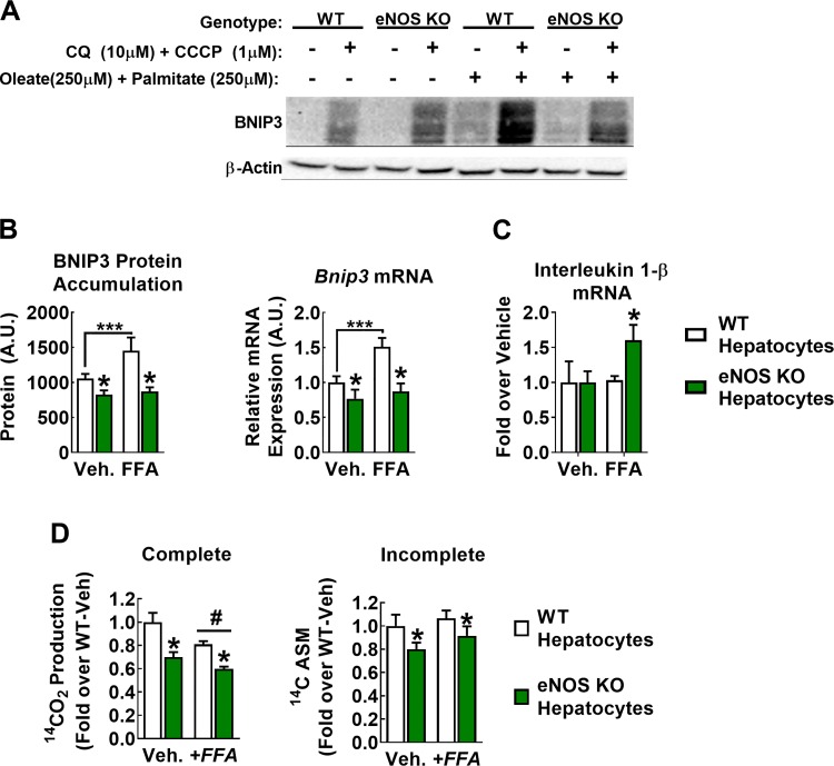
Hepatocellular endothelial NO synthase (eNOS) regulates BCL-2-interacting protein-3 (BNIP3) flux in vitro. A–C: wild-type (WT) and eNOS knockout (KO) hepatocytes were exposed for 24 h with control starvation medium [−free fatty acid (FFA)] or starvation medium conditioned with 500 µM FFA (+FFA; 250 µM palmitate + 250 µM oleate) and treated with 10 µM chloroquine (CQ) and 1 µM CCCP for the final 16 h. A: representative blot of BNIP3 indicating that CQ + CCCP is necessary to induce BNIP3 protein accumulation. B, left: quantification of BNIP3 protein content only in samples that received CQ + CCCP. Right: in a separate experiment, 24-h fatty acid treatment increased BNIP3 mRNA expression in WT but not eNOS KO cells. A.U., arbitrary units; Veh., vehicle. Data are representative of 5–7 biological replicates. #P < 0.05 diet main effect; *P < 0.05 genotype main effect; ***P < 0.05 significant post hoc pairwise comparison indicated by bracket. C: 24-h fatty acid treatment induced mRNA expression of the proinflammatory cytokine interleukin-1β in eNOS KO but not WT hepatocytes. Data are representative of 4 biological replicates. *P < 0.05 paired t-test. D: complete (left) and incomplete (right) [1-14C]palmitate oxidation in WT and eNOS KO hepatocytes. Data are representative of 5–7 biological replicates. #P < 0.05 diet main effect; *P < 0.05 genotype main effect.

