Figure 5.
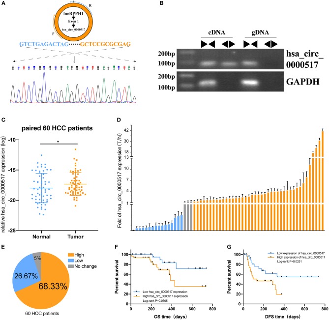
The prognostic value of hsa_circ_0000517 in 60 HCC patients. (A) Sanger sequencing was performed following PCR to validate the head-to-tail splicing of has_circ_0000517 in SNU-387 cells. (B) The validation of RPPH1 circular exon 1, has_circ_0000517. Divergent and convergent primers were used to perform PCR in both cDNA and gDNA. GAPDH was used as a control. (C) The differences in the expression of has_circ_0000517 in 60 tumor and paired normal tissues. *P < 0.05. (D) The Fold Changes (FC) of hsa_circ_0000517 expression in 60 HCC patients (tumor vs. matched non-tumor tissue). Data are represented as the mean ± SD of three independent experiments. Different colors stand for three different groups, including “FC > 1.5,” “FC < 0.67,” and “0.67 < FC < 1.5.” (E) Distribution of three groups in 60 HCC patients. (F) Overall Survival Curve (OS) of has_circ_0000517. (G) Disease-free survival (DFS) curve of has_circ_0000517.

