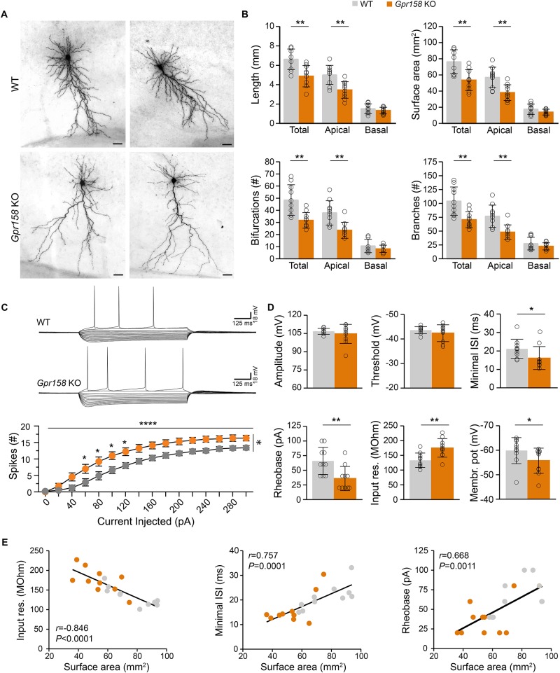
FIGURE 4.
Reduced dendritic architecture supports hyperexcitability of Gpr158 KO CA1 pyramidal neurons. (A) Examples of CA1 pyramidal cells loaded with biocytin during electrophysiological recordings from WT (n = 10 cells from 4 animals) and Gpr158 KO (n = 10 cells from 3 animals) hippocampal slices. Cells were subsequently reconstructed and their basic morphological measures were extracted and analyzed (Online Resource 8). (B) Total dendritic length and surface area were significantly reduced in Gpr158 KO CA1 pyramidals and the effect was specific to the apical compartment. A significant reduction in total and apical but not basal compartments was documented for the number of bifurcations and number of branches. (C,D) For each reconstructed cell, the full action potential profile was generated and analyzed. (C) Example traces of WT and Gpr158 KO action potential profiles generated by current injections in steps of 20 pA. Note that increased number of spikes were observed for all current steps tested in the Gpr158 KO. (D) Action potential amplitude and threshold was unaffected by genotype. Minimum inter spike interval and rheobase were significantly reduced in Gpr158 KO pyramidal cells. In addition, input resistance was increased in Gpr158 KO and the membrane potential was more depolarized in Gpr158 KO pyramidal cells. (E) Significant Pearson correlations (r) and linear regression (Supplementary Figure 4) between cell surface area and input resistance (r = −0.846, P < 0.0001), minimum inter spike interval (r = 0.757, P = 0.0001), and rheobase (r = 0.668, P = 0.0011). Data are presented as mean ± SD; individual data points are indicated. Asterisks indicate significant differences compared with WT cells assessed by Student’s t-test or MWU (Supplementary Tables 4, 5), ∗P ≤ 0.050; ∗∗P ≤ 0.010; ∗∗∗∗P < 0.001.

