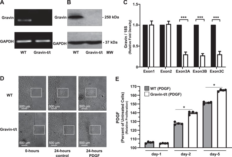
Fig. 8.
Migration and proliferation of aortic vascular smooth muscle cells (VSMCs) isolated from wild-type (WT) and gravin-t/t mice in response to PDGF stimulation. Gravin mRNA expression in aortic VSMCs isolated from WT and gravin-t/t mice was quantified using reverse transcriptase quantitative PCR (A). Gravin protein expression was measured in VSMCs by Western blot from WT and gravin-t/t mice, in which the gravin antibody recognized the monomeric (250 kDa) forms of the gravin protein (B). VSMCs isolated from gravin-t/t mice showed significantly decreased gravin protein expression compared with WT mice. Quantification of the RT-PCR data in A shows that exon 3 of gravin (over three different regions) was significantly decreased in gravin-t/t VSMCs compared with WT VSMCs, as expected (C). Gravin’s exon 1 and exon 2 showed no differences in expression, as expected in this gene-trapped model. Results are presented as mean ± SE. n = 3; ***P < 0.001. Scratch wound assay was used to determine VSMC migration in WT and gravin-t/t mice following 24 h with or without PDGF (10 ng/mL) stimulation (D). Boxed areas emphasize areas of cell migration. Quantification of VSMC proliferation in the presence or absence of PDGF was determined using the MTS colorimetric cell proliferation assay on day 1, 3, and 5 after PDGF stimulation (E). Results are presented as the mean ± SE. n = 4; *P < 0.005. Comparisons between two groups were determined by an unpaired two-tailed Student’s t test, and comparisons between multiple groups were determined by one-way ANOVA followed by post hoc Tukey test. MM, molecular mass.

