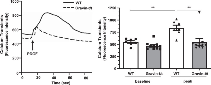
Fig. 9.
Intracellular Ca2+ transient in vascular smooth muscle cells (VSMCs) isolated from wild-type (WT) and gravin-t/t mice in response to PDGF stimulation. Intracellular Ca2+ transients tracing (left) recorded from aortic VSMCs isolated from WT and gravin-t/t mice before and after PDGF (10 ng/mL) stimulation. Quantitative analysis of fluorescence intensity of Ca2+ transients (right) demonstrated that absence of gravin-mediated signaling inhibited PDGF-induced Ca2+ release. Results are presented as the mean ± SE. n = 7 (WT ND), n = 7 [WT high-fat diet (HFD)], n = 10 (Gravin-t/t ND), and n = 10 (Gravin-t/t HFD). **P < 0.01. Comparisons between two groups were determined by an unpaired two-tailed Student’s t test, and comparisons between multiple groups were determined by one-way ANOVA followed by post hoc Tukey test.

