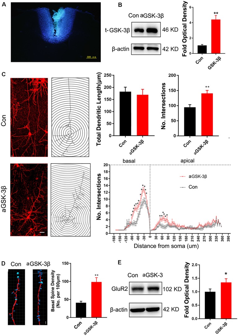
Figure 4.
Effects of over-expressing constitutive active GSK-3β (aGSK-3β) in Shank3b−/− ACC on the development of dendrites and spines. (A) Hoescht33342 staining of injection site at 24 h after virus injection in Shank3b−/− mice of 4 weeks old. (B) Western-blotting of t-GSK-3β in ACC at 3 weeks after injecting AAV expressing aGSK-3β. (C) Sholl analysis of Golgi images of pyramidal neurons in control and aGSK-3β treated Shank3b−/− ACC. Notice the increase of dendritic intersections, and particularly basal branches in the aGSK-3β treated Shank3b−/− neurons. (D) Imaris analysis of spines along basal dendrites of control and aGSK-3β treated Shank3b−/− neurons. Notice the increase of spine density by aGSK-3β. Values represent mean ± SEM. *P < 0.05. **P < 0.01. Bar = 20 μm (C) and 2 μm (D). N = 3 mice per group (B), and 6 mice per group (C–E). Student’s t-test (A,D,E). Two-way ANOVA (B).

