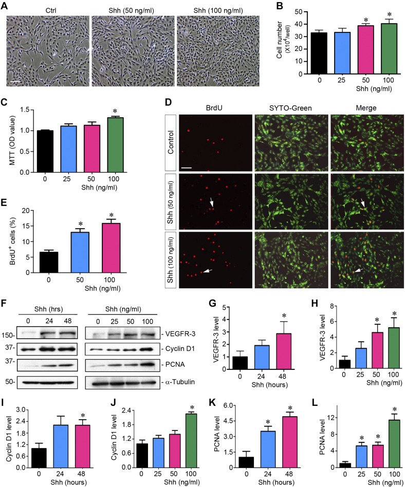
Fig. 3.
Sonic hedgehog (Shh) promotes lymphatic endothelial cell proliferation in vitro. A: representative micrographs showing phase-contrast images of human dermal lymphatic endothelial cells (HDLECs) after incubation with recombinant Shh protein. HDLECs were incubated with Shh for 48 h at different concentrations as indicated. Scale bar = 30 µm. B: Shh promotes HDLEC proliferation in a dose-dependent manner. Cell numbers were counted at 48 h after incubation with Shh and are shown. Data were obtained from three independent experiments. *P < 0.05 vs. controls (Ctrl; n = 3). C: graphic presentation showing that Shh promoted HDLEC density assessed by a colorimetric MTT assay. *P < 0.05 vs. controls (n = 3). OD, optical density. D: representative micrographs showing that Shh promoted HDLEC DNA synthesis and entering the S phase as shown by bromodeoxyuridine (BrdU) incorporation. Cells were immunostained with anti-BrdU antibody (red) at 48 h after incubation with different concentrations of Shh. SYTO-Green (green) was used to visualize nuclei. Scale bar = 30 µm. E: quantitative determination of the percentage of BrdU+ cells after Shh treatment. *P < 0.05 vs. controls (n = 3). F: Western blots showing that Shh promoted the expression of VEGFR-3 and proliferation-related genes. HDLECs were incubated with different concentration of Shh for 48 h or Shh (50 ng/ml) with various durations. G–L: cell lysates were subjected to Western blot analyses for VEGFR-3 (G and H), cyclin D1 (I and J), and proliferating cell nuclear antigen (PCNA; K and L), respectively. *P < 0.05 vs. controls (n = 3).

