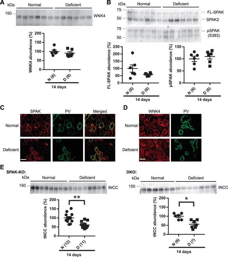
Fig. 2.
Long-term (14 days) dietary Mg2+ restriction did not affect with-no-lysine (K) kinase-4 (WNK4) or STE20/SPS-1-related proline/alanine-rich kinase (SPAK). A: Western blot analysis of whole kidney lysates did not show any significant difference in the abundance of WNK4 between normal diet (N) and Mg2+-deficient (D) diet groups (two-tailed unpaired t-test). B: full-length SPAK (FL-SPAK) and phosphorylated SPAK (pSPAK) abundances also did not differ between normal and Mg2+-deficient diets (two-tailed unpaired t-test). C: immunofluorescence for SPAK (red) showed similar localization in normal and Mg2+-deficient diets. Colocalization with parvalbumin (PV; green) showed that SPAK was localized along the distal convoluted tubule (DCT). D: WNK4 (red) localization did not differ between normal diet and Mg2+-deficient diet. PV (green) was used to identify DCT in the same section. Note that on normal diet and low-Mg2+ diet, WNK4 signal is weak and diffuse along the DCT and DCT2/connecting segment (CNT), in contrast to what is observed after low-K+ diet (62) or genetic activation of the WNK4-SPAK pathway (22), so the images were not overlaid. Results are representative of 5 independent experiments. Scale bars in C and D = 40 μm. E: dietary Mg2+ restriction downregulated total Na+-Cl− cotransporter (tNCC) abundance in SPAK knockout (KO) mice. tNCC abundance was also significantly reduced in SPAK and oxidative stress-response kinase-1 (OSR1) double-KO (DKO) mice on Mg2+-deficient diet compared with normal diet (two-tailed unpaired t-test). For blot quantification, densitometric values were normalized using Coomassie-stained gels. Values are means ± SE; values in parentheses indicate n values. *P < 0.01; **P < 0.001.

