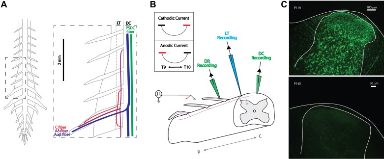
Fig. 2.
Ex vivo adult mouse preparation: experimental setup and viability. A: schematic of gross anatomy of spinal cord from dorsal surface and simplified organization of axon fiber populations subject to recruitment by spinal cord stimulation (SCS; inset). Axonal recruitment with SCS was assessed in dorsal roots (DR), dorsal column (DC), and Lissauer tract (LT). Whereas the DC predominately carries nonnociceptive sensory information, the neighboring LT contains projection from nociception transmitting primary afferents. PSDC, postsynaptic dorsal column. B: schematic of general experimental paradigm. Two electrodes (~1.5 mm apart) are placed on or above the medial DC in a region between T9 and L3. This is used to simulate a bipolar SCS system. Responses to SCS are recorded via glass suction electrodes on the DC and DRs and by a microelectrode on LT. Inset: orientation of bipole (cathode, black; anode, red) and direction of current flow for cathodic and anodic stimulation used for experimental studies. R and C denote rostral and caudal, respectively. C: postexperimentation Nissl staining revealed persistent cellular viability 8 h after cord isolation (top) but dramatic deterioration when superfused for 2 h in the absence of oxygenation (bottom).

