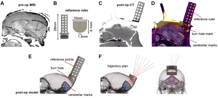
Fig. 2.
Development of a chamber-based geometric model to define electrode trajectories and specify location of the burr hole. A: preoperative (pre-op) MRI image used for identifying the desired regions of interest in the cerebellum. B: reference axis ruler (1.104 g) that was inserted into the base chamber before postoperative computer tomography (post-op CT) imaging. C: post-op CT image after surgical installation of the head post, base chamber, and reference ruler. Although the reference ruler is clearly visible, the titanium head post and chamber have produced significant artifacts. D: coregistered pre-op CT, post-op CT, and pre-op MRI. Markers identify points on the reference axis and points in the cerebellum. A mark identifies the burr hole location. E: a 3-dimensional (3D) model that has coregistered the skull, cerebellum, chamber, and reference ruler. F: using the 3D model, we drew trajectories that began at points of interest in the cerebellum, converged on a single 1.5-mm-diameter burr hole on the skull, and then diverged beyond the chamber as cylinders within the guidance tool. These trajectories represented desired electrode paths.

