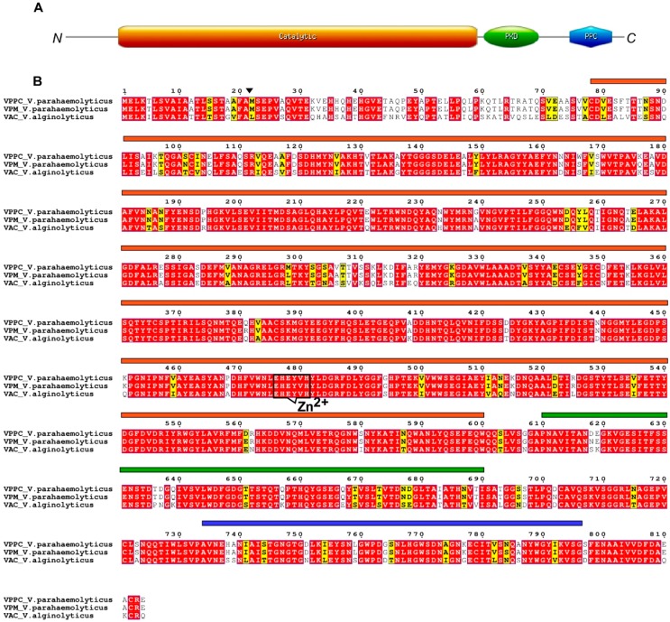
Figure 4.
Schematic diagram of M9A class III collagenases from V. parahaemolyticus and V. alginolyticus. (A) Collagenases possess a N-terminal signal peptide and a proteolytic activation site marked by an arrow. The catalytic, the PKD-like and PPC domains are showed in orange, green and blue, respectively. (B) Sequence alignment of collagenases from V. parahaemolyticus and V. alginolyticus. Amino acids forming the zinc-binding motif are shown. Similar residues are written in black bold characters and boxed in yellow, whereas conserved residues are in white bold characters and boxed in red. The alignment was performed with T-Coffee. The sequence numbering on the top refers to the alignment.

