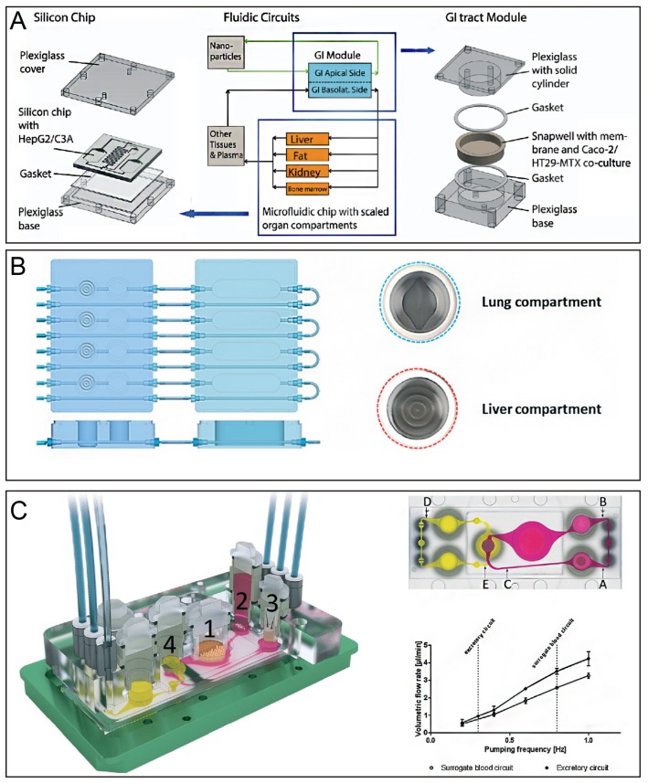
Figure 5.
(A) Gastrointestinal tract and liver tissue system construct by combining the human intestinal epithelium and liver represented by coculture of enterocytes (Caco-2) with mucin-producing cells (TH29-MTX) and HepG2/C3A cells. Reproduced with permission from [109]. (B) Lung/liver-on-a-chip, in which liver spheroids were connected in a single circuit and normal human bronchial epithelial cells were cultured at the air-liquid interface. Reproduced with permission from [98]. (C) The microfluidic four-organ-chip device. (i) 3D view of the device comprising two polycarbonate cover plates, a PDMS-glass chip accommodating a surrogate blood flow circuit (pink), and an excretory flow circuit (yellow). Numbers represent the four tissue culture compartments for the intestines (1), liver (2), skin (3), and kidney (4). (ii) Central cross-section of each tissue culture compartment aligned along the interconnecting microchannel. (iii) Average volumetric flow rate plotted against the pumping frequency of the flow circuit. Reproduced with permission from [113].

