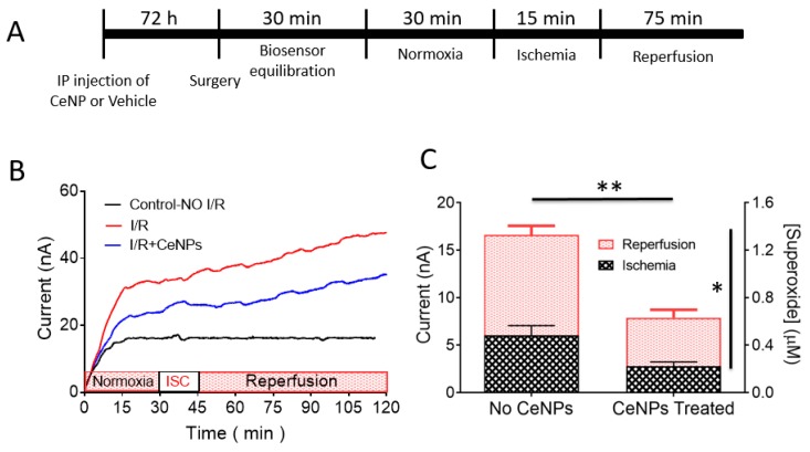
Figure 6.
(A) Experimental timeline (not drawn to scale) for in vivo monitoring of superoxide during ischemia (right common carotid artery occlusion) and reperfusion in rats. (B) In vivo amperometric current–time response of the Cyt C microbiosensor showing continuous monitoring of superoxide anion radical in rat hippocampus at an applied biosensor potential of +0.15 V vs. Ag/AgCl. Representative amperograms for the control with no ischemia (black), ischemia and reperfusion (red) and ischemia and reperfusion in the CeNPs-treated rats (blue) are displayed. Ischemia (ISC) was induced for 15 min (30–45 min time point) and followed by reperfusion for 75 min (45–120 min time point). (C) Quantification of superoxide levels in vivo in rat brain hippocampus during ischemia and reperfusion. Bar graphs show the contribution of superoxide during ischemia (black hatched area) and reperfusion (red hatched area) in both CeNP-treated (n = 9) and vehicle control (no CeNPs) (n = 7) animals. Values are expressed as mean ± SEM. A two-way ANOVA revealed two main effects: a significant reduction (** p < 0.0001) in the total concentration of superoxide (ischemia plus reperfusion) in CeNPs-treated animals compared to controls (no CeNPs), and the SO concentration was significantly greater during reperfusion compared to ischemia (* p = 0.0015).

