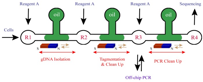
Figure 7.
Schematic of microfluidic workflow [81]. Water or aqueous buffer exists in each reservoir (R1–R4). Cells are added in R1 and incubated, followed by adding of reagent A (magnetic beads and binding buffer). The beads binding with DNA are moved to R2 and then DNA is eluted from the beads in reaction buffer. DNA fragmentation and adapter ligation reaction are performed in R2. After reagent A is added and beads are moved to R3, DNA is eluted from the beads into water. DNA is transferred for off-chip PCR amplification and then moved back to R3. With Reagent A added in R3 and beads moved to R4, purified DNA is prepared for sequencing.

