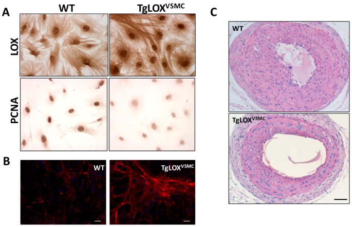
Figure 5.
LOX transgenesis limits VSMC proliferation and vascular remodeling. (A) Immunocytochemical analysis of LOX and PCNA, markers of cell proliferation, in aortic VSMC from TgLOXVSMC and WT mice. (B) Collagen type-I deposition (in red) by VSMC from TgLOXVSMC and WT mice visualized by confocal immunofluorescence in nonpermeabilized cells, as described [39]. Cells were incubated with a COL1A1 specific antibody (NB600-408; Novus Biologicals, UK) and nuclei were stained with DAPI. As observed, LOX transgenesis induces the deposition of a thicker and more organized collagen network (Bar: 25 µm). (C) TgLOXVSMC and WT mice were subjected to left common carotid artery ligation. Injured arteries were harvested 21 days after surgery, fixed in 4% paraformaldehyde, and embedded in paraffin. Cross-sections at 1.5 mm from the ligation site were stained with hematoxylin and eosin, as described [62,63]. This staining evidenced that LOX overexpression reduces neointimal growth (Bar: 100 µm).

