Figure 6.
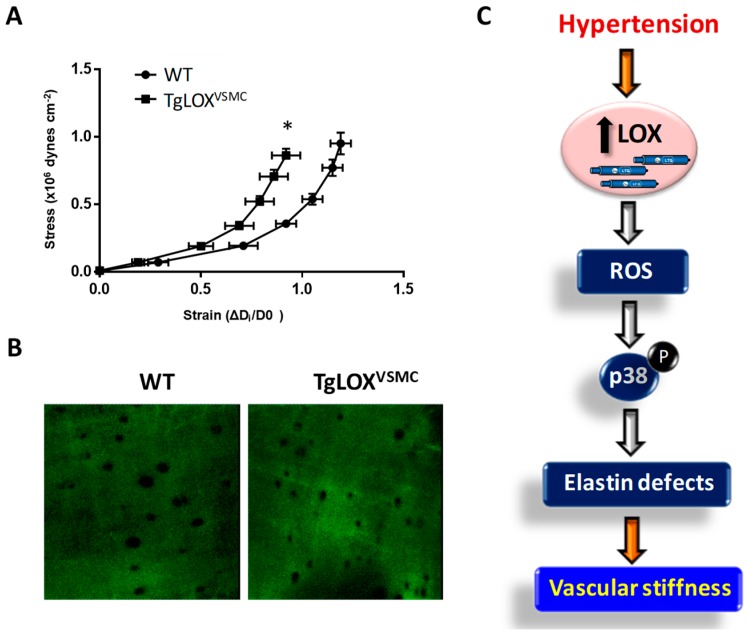
Contribution of LOX to oxidative stress and vascular stiffness in hypertension. (A) The structural and mechanical properties of first-order mesenteric arteries were studied with a pressure myograph (Danish Myo Tech, Aarhus, Denmark) as previously described [78,79]. Stress–strain curves were obtained in arteries from WT and TgLOXVSMC mice. Vascular stiffness was determined from the stress–strain relationship which is nonlinear; therefore, we obtained the incremental elastic modulus (Einc) by determining the slope of the stress–strain curve of individual animals. Einc values were (mean ± SEM) WT: 4.11 ± 0.12 (n = 6); LOX: 5.17 ± 0.27 (n = 8). *p < 0.05 vs. WT by unpaired student t-test. For simplification, statistical analysis is shown in the stress–strain curves. (B) The elastin organization within the internal elastic lamina was studied in segments of mesenteric arteries from TgLOXVSMC and WT mice using fluorescence confocal microscopy based on the autofluorescent properties of elastin (ex: 488 nm; em: 500–560 nm), as previously described [78,79]. Serial optical sections from the adventitia to the lumen (z step = 0.5 µm) were captured with a X63 oil objective, by using the 488 nm line of the confocal microscope. (image size 53 × 53 µm). (C) Working model showing that LOX induced-oxidative stress alters elastin structure and promotes vascular stiffness in hypertension.

