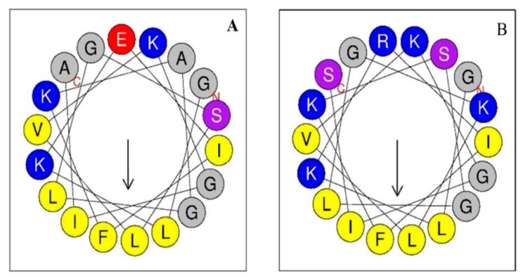
Figure 1.
Wheel projections of the first 18 residues of the sequence of each peptide. Alyteserin-1c peptide (+2) (A); peptide +5 (B). The hydrophobic amino acids are yellow, and the charged amino acids are blue (positive charge) or red (negative charge). The polar amino acids are purple and those in between are grey.

