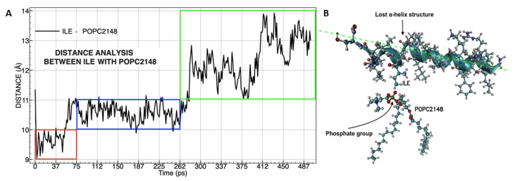
Figure 9.
Molecular distances analysis between isoleucine (ILE17) with phosphatidylcholine (A), the color frames represent three times of affinity during molecular dynamics. Molecular representation was taken during peptide insertion into the membrane (B). The specific forces of van der Waals called dipole–dipole forces are represented between the phosphate group (polar) in POPC and the side chain (non-polar) of ILE (C-H----O-P). The green dotted line shows the change of the spatial position of peptide +5 that causes the loss of α-helix structure and displacement of the LVKGIS sequence.

