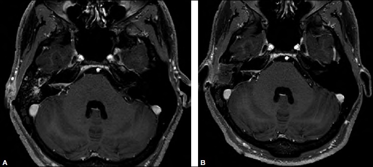
Fig. 4.

MRI, T1 sequence after gadolinium infusion, axial view, before surgery: clear reduction of the enhancement of the internal auditory canal; no more enhancement is visible at the level of the inner ear; B) control MRI, T1 sequence after gadolinium infusion, axial view, 3 months after the surgery: external and middle ear are packed with fat tissue; further reduction of the enhancement of the internal auditory canal.
