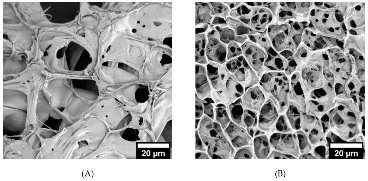
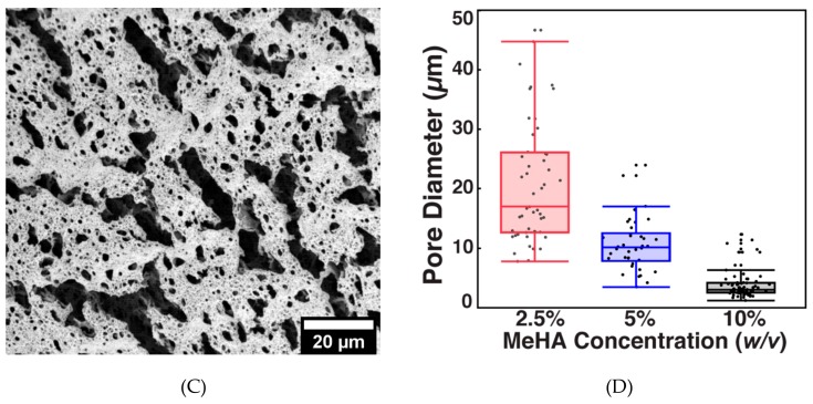
Figure 3.
Pore size analysis of crosslinked x-MeHA hydrogels. (A) 2.5% w/v, (B) 5% w/v, and (C) 10% w/v hydrogels were crosslinked and lyophilized and then analyzed under SEM. (D) Pore diameters analyzed via ImageJ and JMP. The 5% and 10% w/v hydrogels had significantly smaller pore diameters (two-sample t-tests) compared to the 2.5% w/v hydrogels. Scale bar is 20 µm.


