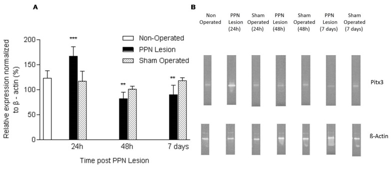
Figure 2.
Effect of pedunculopontine nucleus (PPN) neurotoxic lesion on the paired-like homeodomain transcription factor 3 (Pitx3) mRNA expression in nigral tissue. (A) Comparison between experimental groups 24 h (F (2, 14) = 11.06 p < 0.001), 48 h (F (2, 17) = 7.22 p < 0.01) and seven days (F (2, 15) = 7.65 p < 0.01) after PPN lesion. The first bar (white bar) represents the non-operated group. This group is common for the ANOVA statistical analysis, which was carried out among non-operated, PPN lesion and sham-operated groups for each time post lesion separately. The asterisks correspond to statistical differences between PPN lesion and both control groups (non-operated and sham operated). (B) Agarose/ethidium bromide gel electrophoresis bands representatives of the semi-quantitative RT-PCR study for Pitx3 mRNA expression. *** p < 0.001; ** p < 0.01.

