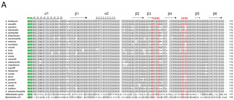
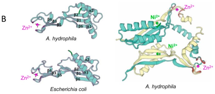
Figure 1.
(A) Sequence alignment of the in silico-translated amino acid sequences of HypA proteins from 36 Aeromonas species, E. coli and H. pylori. The alignment was constructed with MegAlign. The MHE correspond to the motif of the Nickel binding domain (green) and CxxCnCPxP to the Zinc binding domain (red). The characteristic α-helices (wave lines) and a β-sheet (arrows) are represented. (B) Predicted monomeric and dimeric structure of HypA proteins from A. hydrophila type strain and E. coli constructed with Swiss Model online tool, the α-helices and the stranded β-sheet motifs are indicated.


