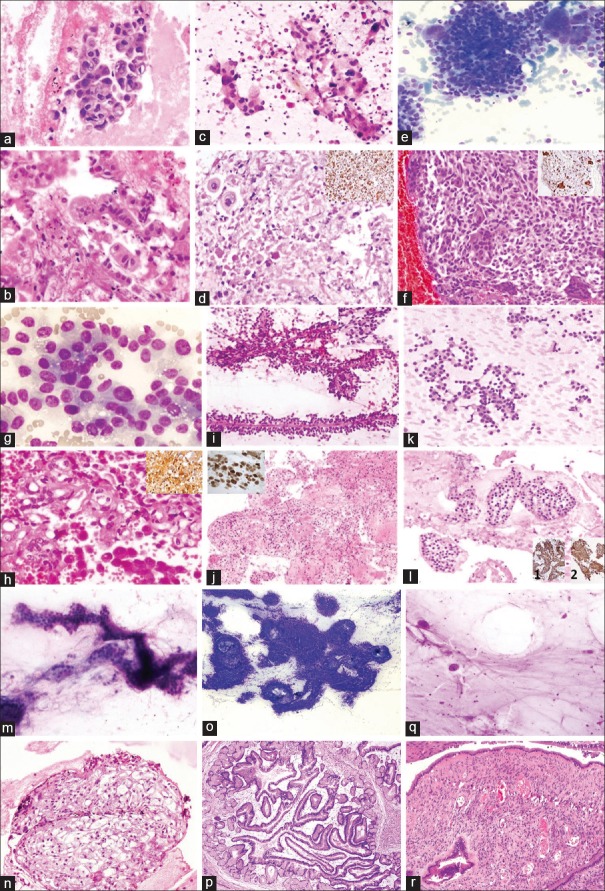
Figure 4.
Adenocarcinoma: Cytology smear [a; H and E; ×200] and CB histology [b; H and E; ×200]. Adeno-Squamous Carcinoma: Smear [c; H and E; ×200] and CB histology [d; H and E; ×200]. IHC for CK7 is negative in squamous component [Inset; ×200]. Undifferentiated Pancreatic Carcinoma with Osteoclastic Giant Cells [e: cytology smear; MGG stain; ×200], [f: CB histology; H and E; ×200], giant cells are positive for CD68 [Inset; ×200]. Acinic Cell Carcinoma: Cytology smear [g; MGG stain; ×400] and CB histology [h; H and E; ×200], IHC for chymotrypsin is positive [Inset; ×200]. Solid Pseudopapillary Epithelial Neoplasm: Cytology smear [i; H and E; ×200] and CB histology [j; H and E; ×200] IHC shows nuclear positivity for beta catenin [Inset; ×200]. Neuroendocrine Tumor: Smear [k; MGG stain; ×200] and CB histology [l; H and E; ×200]. IHC shows cytoplasmic positivity for synaptophysin [1; ×200] and chromatogranin [2; ×200]. Serous Cystadenoma: Cytology [m; MGG stain; ×200], CB histology [n; H and E; ×200]. Intraductal Papillary Mucinous Neoplasm: Cytology [o; MGG stain; 200], Histology section [p; H and E; ×200]. Mucinous Cystadenoma: Cytology [q; MGG stain; ×200] and Histology [r; H and E; ×200]

