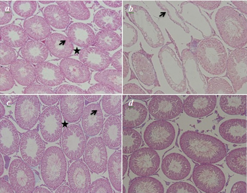
Figure 2.

Histopathological analysis of the p-NP- and GTE-treated rats using the Light microscopic study. (a) and (c) the control and the GTE groups manifesting a normal feature of the seminiferous epithelium (arrow) and interstitial tissue (star). (b) the p-NP group revealing a markable shrinkage of empty seminiferous tubules (arrow). (d) the p-NP + GTE group – the undesired changes are ameliorated toward the normal structure (Magnification: 100).
