
Figure 1.
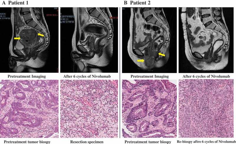
Radiologic and pathological response to neoadjuvant treatment with nivolumab.
Panel A shows pelvic MRI of patient 1 with stage cT4bN2M0 rectal cancer before and after the administration of nivolumab. In the upper row, the pretreatment scan shows a large mass in the upper middle of rectum was adhered to left seminal vesicle gland, the posterior wall of the bladder and the presacral space (arrow). The distance from inferior margin of tumor to the anal verge was 6.4 cm and the tumor length was 8.0 cm. A scan performed before surgery shows greatly shrinkage of the tumor. In the lower row, shown are representative sections of tumor specimens obtained from patient 1 before the administration of nivolumab (left) and after the administration (right) (hematoxylin and eosin staining). This patient had 100% pathological regression of the primary tumor. Panel B shows Pelvic MRI of patient 2 with stage cT4bN2M0 rectal cancer, who received six cycles of nivolumab as neoadjuvant treatment. In the upper row, before the infusion of nivolumab (left), the tumor grows out of intestine and invaded the vaginal inferior wall, the perineal shallow area, the posterior wall of the vaginal vestibule, the anal canal, and the right levator ani muscle (arrow). After six cycles of nivolumab, no extraluminal infiltration was observed. The lesion was significantly shrunk and scarring. In the lower row, shown are representative sections of tumor specimens from patient 2 before the administration of nivolumab (left) and after administration (right) (hematoxylin and eosin staining). Tumor cells are present throughout the pretreatment specimen, whereas in the post-treatment biopsy specimen, there were no viable cancer cells.