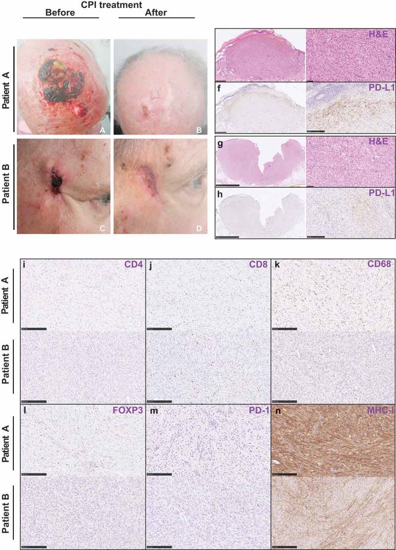
Figure 1.

Clinical images and histology of patient A and patient B.
Clinical images of patient A (a, b) and patient B (c, d) before and after immunotherapy. Hematoxylin-Eosin stains (e, g) and immunohistochemical pictures of various markers of immune cells for patient A and patient B (f, h, i-n).
