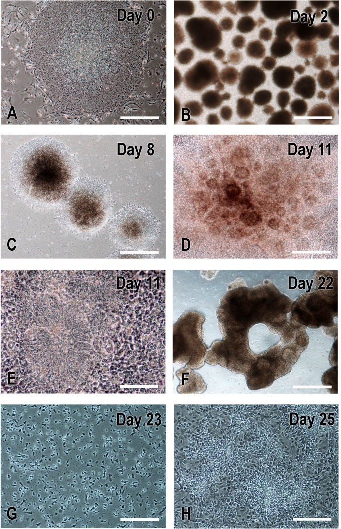
Fig 3. Critical steps in neural differentiation of human ES cells (Day 0) to NPs (Day 25).
On Day 0, H9 ES cell colonies (A) were detached using collagenase to form embryoid bodies (B; EBs on Day 2) that were further cultured in EB media with dorsomorphin and A-83. Beginning at day 4, medium was changed to the one promoting NP cell differentiation that was used to feed every other day to Day 22. On Day 7, EBs were attached to matrigel-coated plate; neural rosette structures were evident at day 8 (C) and became prominent on day 11 (D-E). On day 22, colonies with rosettes were detached manually to form neurospheres (F). On day 23, neurospheres were dissociated into single cells with accutase, plated on matrigel-coated plate, and fed with NP expansion media till they became confluent (G-H). Cells were passed every 4–5 days. Days 36 NPs were used for viral transduction and Day 41 NPs for transplantation. Scale bars: A-C, F, 500μm; D and G-H, 200μm; E, 100μm.

