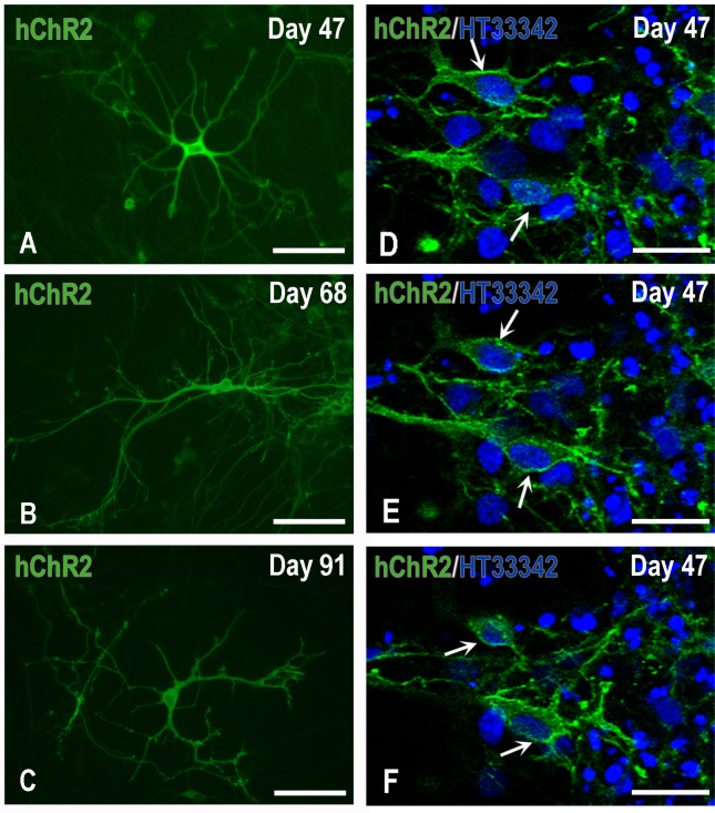
Fig 5. Expression and localization of hChR2 in H9-derived hNPs.
Panels A-C depict typical neurons at various days of maturation in vitro: H9 hNPs, photographed here on day 47 in culture, had been transduced on Day 34 by lentivirus carrying hChR2 at moi 13. Human ChR2-YFP expression started to appear two-four days after transduction and progressively spread over the perikaryon and processes of maturing neurons. Panels D-F demonstrate the fine localization of hChR2 (YFP fluorescence), predominantly to the membranes of representative transduced nerve cells (arrows), seven days after transduction, by confocal microscopy. Scale bars: A-C, 100μm; D-F, 50μm.

