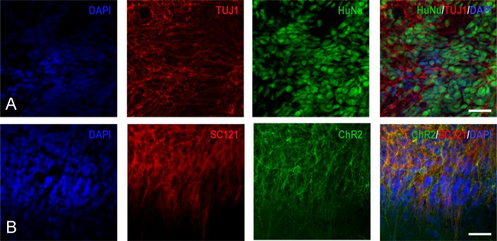
Fig 8. Series of confocal images indicating the differentiation of transplant-derived cells into early neurons with hChR2 expression.
(A) These four panels indicate that a large percentage of transplant-derived HuNu+ cells (green) are also immunoreactive with antibodies against class III β-tubulin epitope TUJ1 (red). Blue nuclei are stained with DAPI. (B) These panels indicate that the majority of transplant-derived epitope SC121+ positive cells (red) also fluoresce in the green spectrum, which is indication of hChR2 expression. Cell nuclei are stained with DAPI (blue). Scale bars: 200μm.

