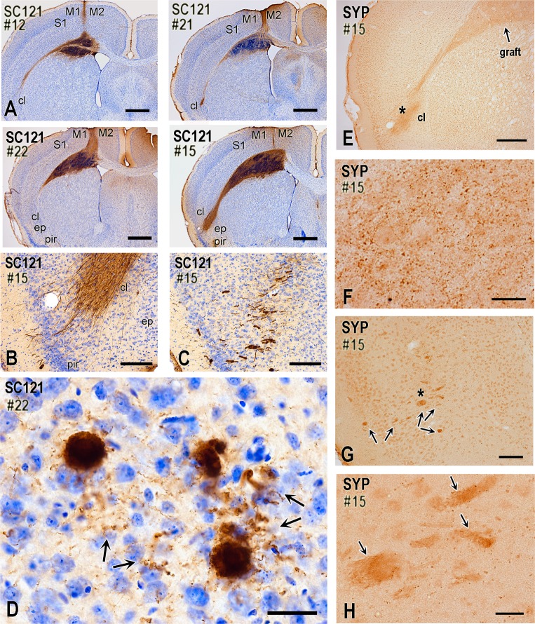
Fig 9. Neuronal differentiation of ChR2-transduced and non-transduced hNP transplants and pathfinding of axons from their neuronal progenies two months after transplantation (mice).
(A) These four panels are from representative cases visualized with IHC for the human-cell specific epitope SC121 and illustrate a consistent engraftment of hNPs in the primary motor area at a coronal level corresponding to caudal septum. Transplant engages deep cortical layers, medial corpus callosum, and dorsal neostriatum. In most cases, the transplantation needle track is also evident. Panels also show that SC121+ transplant-derived axons project along the fibers of corpus callosum and exit at the level of claustrum. Transplants in #21 and #22 contain transduced hNPs, whereas in #12 and #15 contain non-transduced cells. All cases are from injured mice. (B-D) SC121 immunohistochemical preparations illustrating the propensity of transplant-derived axons to exit the lateral corpus callosum at the level of the claustrum and then course in deep insular/piriform cortex. Case 15 involves non-transduced NPs. There is a striking concentration of axons at the claustrum, and an occasional further advancement towards the insular and piriform cortex. In some cases, bundles of axons extend all the way to layer II of piriform/insular cortex (B), commonly following a course oblique or vertical to the plane of view (C). Panel D shows that axon bundles branch into numerous single axons with boutons (arrows) in the deep insular/piriform cortex (Case 22, transduced NPs). (E-H) These human synaptophysin-immunostained preparations show the formation of early synaptic fields by transplant-derived neurons. Low-level synaptophysin expression often extends from the site of the transplant all the way to the lateral corpus callosum and insular/piriform area, with an apparent terminal field at the claustrum/piriform cortex (E). With higher magnification, terminal field contains putative synaptic boutons (F; photograph is taken from the claustral/piriform area indicated with asterisk on panel E). Bundles of axons that course obliquely or vertically to the coronal plane as in panels C-D also express diffuse synaptophysin immunoreactivity (G; bundles are indicated with arrows). Synaptophysin+ boutons surround such bundles (H; panel is a magnification of area indicated with an asterisk on panel G). M1, primary motor cortex M2, secondary motor cortex; S1, primary somatosensory cortex; cl, claustrum; ep, endopiriform nucleus; pir, piriform cortex; SYP, synaptophysin. Scale bars: A, 800μm; B-C, 200μm; D, 30μm; E, 400μm; F-H, 20μm; G, 100μm.

