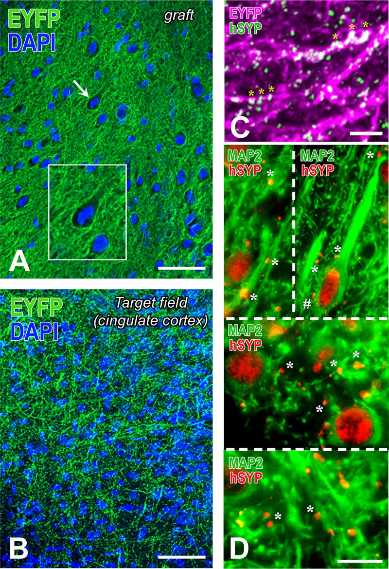
Fig 10. Differentiation of hChR2-hNP transplants in rat motor cortex and patterns of innervation of host neurons.

(A-B) These YFP-immunostained preparations show the distinct features of hChR2+ neural tissue in the transplant (A) versus a hChR2+ terminal field in cingulate cortex (B). YFP expression in (A) is restricted to the membrane of the neurons and their processes. The latter form a dense neuropil with webs of processes filling the space between neuronal cell bodies. Some of these hChR2-hNP-derived neurons, for example the one indicated with the arrow and enlarged in the inset have cortical features. The terminal field of hChR2-hNP-derived neurons in cingulate cortex (B) has a very different appearance than the “neuropil” in the transplant and it obeys the cytoarchitecture of host cortex. No perikaryal-type staining is encountered, evidence that graft-derived neurons have not migrated into host cortex. (C) This dually stained preparation for two transplant-selective neuronal markers (YFP immunoreactivity for hChR2 and human synaptophysin immunoreactivity) is from layer II of host cingulate cortex and demonstrates both the dense terminal field and the extensive colocalization of the two markers in transplant-derived axons and their processes (asterisks; double labeling is white here). A larger panel showing more of this terminal field is in S6 Fig. (D) These dually stained preparations from host motor and cingulate cortex with MAP2 for neurons and human synaptophysin for transplant-derived terminals show a very large number of terminals apposing dendrites and dendritic branches of host neurons in layer 2–3 of motor cortex (top left), layer 5 of motor cortex (top right), deep layer 2–3 of cingulate cortex (middle) and superficial layer 2–3 of cingulate cortex (bottom). In many cases, dendrites are sectioned transversely. There are occasional terminals on the perikarya of large neurons (see an example on a pyramidal neuron labeled with #). Scale bars: A-B, 50μm; C, 10μm; D, 20μm.
