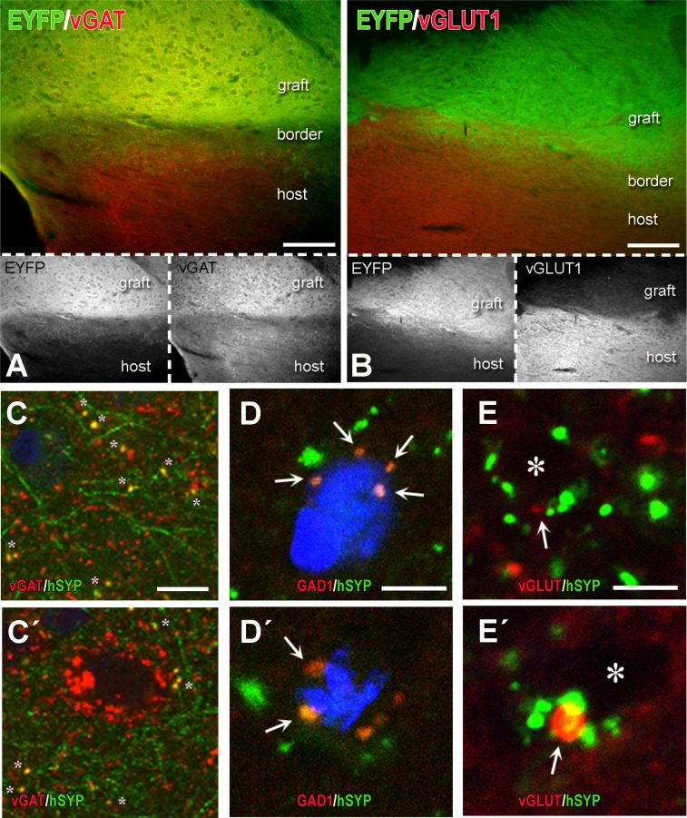
Fig 11. Partial characterization of neurotransmitter identity of differentiated hChR2-hNP transplants and their terminals in host brain.
(A-B) These are dually immunostained preparations for YFP (hChR2 marker to label the transplant and transplant-derived structures) and either vGAT, a presynaptic marker of GABAergic neurotransmission (A) or vGLUT1, a presynaptic marker of glutamatergic neurotransmission (B). Color panels show both transplant and neurotransmission marker immunoreactivities, whereas black and white panels underneath the color ones illustrate transplant and neurotransmitter markers separately. Note the dense GABAergic neurotransmission in the graft (orange neuropil in A) as contrasted with the very low glutamate marker immunoreactivity (B). (C-E´) Representative images illustrating the GABAergic or glutamatergic differentiation of transplant-derived human synaptophysin+ (hSYP+) terminals in rat motor cortex. Differentiation was assessed with IHC for GABAergic markers such as vGAT (C-C´) or GAD1 (D-D´) and glutamatergic markers such as a mixture of antibodies for vGLUT 1 and 2 (vGLUT) (E-E´). Images in C-C´ are taken at a lower magnification than D-E´. There are multiple vGAT+ or GAD1+ transplant-derived hSYP+ terminals (yellow color), most of them on non-identifiable host structures, probably dendrites (asterisks in C-C´) but quite a few also on somata (double asterisk on C´ as part of a basket-type GABAergic innervation of a rat cortical neuron; arrows on D-D´). At the contrary, we were not able to find any colocalization of vGLUT in hSYP+ terminals (D-D´); even when there is double labeling as in E´, the relationship is that of juxtaposition, not colocalization. Asterisks on E-E´ are unlabeled rat (host) structures, probably dendrites. Scale bars: A-B, 100μm; C-E, 10μm.

