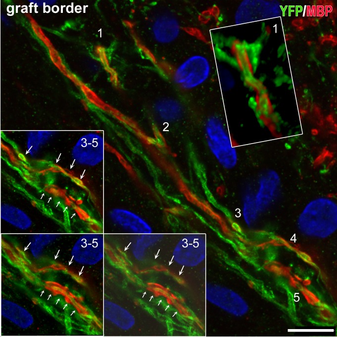
Fig 12. Myelinated human axons in dually immunostained preparations with YFP antibodies (for hChR2+ axons) MBP (for myelin) through the border of a transplant.
Single optical sections are taken with a confocal microscope. Human ChR2-hNP-derived axons are in green and myelin in red. At least a third of these axons is myelinated. Inset on top right is a 3D rendition of profile 1 in main frame from z-stack. Insets on bottom left are optical sections of profiles 3–5. Note the non-continuous myelination in the form of serial MBP+ “cuffs”. Graft is on the left. Rat corpus callosum is on top right. Scale bar: 10μm.

