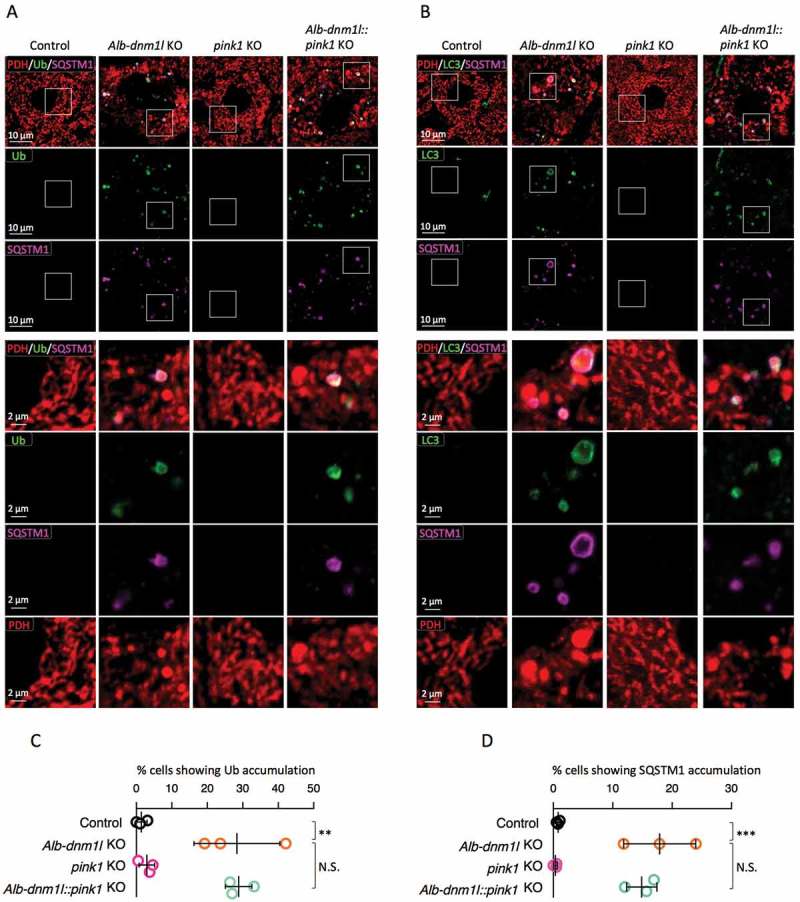
Figure 2.

Mitochondria undergo ubiquitination in the absence of PINK1. (A and B) Frozen liver sections were examined by immunofluorescence microscopy with antibodies against ubiquitin, SQSTM1, and PDH (A) and LC3, SQSTM1, and PDH (B). Lower panels are magnified images of boxed regions. Bars in higher panels: 10 µm. Bars in lower panels: 2 µm. (C and D) Quantification of cells showing the accumulation of ubiquitin (C) and SQSTM1 (D) on mitochondria. Values are average ± SD (n = 3 mice). Statistical analysis was performed using one-way ANOVA followed by the post-hoc Tukey test: *p < 0.05, **p < 0.01, ***p < 0.001.
