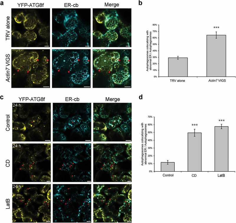
Figure 7.

Punctate ER remnants colocalize with autophagic structures in Actin7-silenced plants and 24 h anti-microfilament drug-treated leaves. (a) Representative confocal images showing colocalizations of ER remnants with YFP-ATG8f-labeled autophagic structures (yellow) in Actin7-silenced plants and control plants. ER network was labeled by ER-cb (cyan). Red arrowheads indicate the colocalized structures. Scale bars: 10 μm. (b) Ratio of autophagic structures colocalized with punctate ER remnants to total autophagic structures in Actin7-silenced plants. More than 150 cells were quantified in each treatment. Values are means ± SE from 3 independent experiments. Student’s t test was performed to indicate significant difference (*** p < 0.001). (c) Representative confocal images showing colocalizations of ER remnants with YFP-ATG8f-labeled autophagic structures (yellow) in leaves treated with 20 μM CD or 25 μM LatB for 24 h. Red arrowheads indicate the colocalized structures. Scale bars: 10 μm. (d) Ratio of autophagic structures colocalized with punctate ER remnants to total autophagic structures in drug-treated leaves. More than 100 cells were quantified in each treatment. Values are means ± SE from 3 independent experiments. Student’s t test was performed to indicate significant difference (*** p < 0.001).
