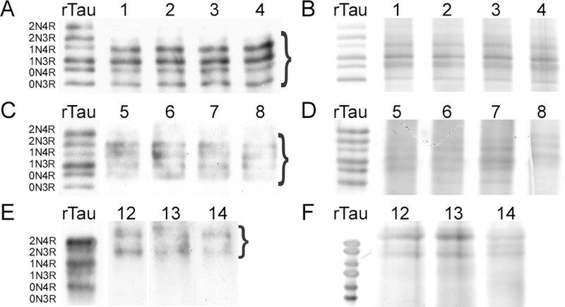
Fig. 3.
SDS-PAGE analysis of tau proteins isolated from cognitively normal middle age (A,B) cognitively normal elderly (C,D) and AD (E,F) brain samples. Lanes correspond to case numbers summarized in Table 1. Electrophoretic migration of all samples is shown relative to a ladder composed of all six human tau isoforms (rTau). (A, C, E) Gels were subjected to immunoblot analysis with monoclonal antibody Tau5. (B, D, F) Gels were stained with silver. Bands excised for targeted metabolomics analysis are marked by brackets.

