Figure 3.
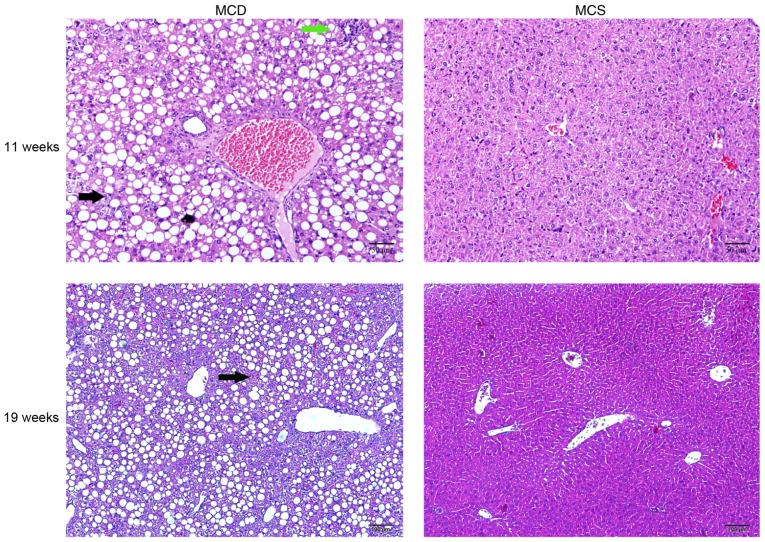
NAFLD was established by feeding an MCD diet to C57BL/6J mice. H&E staining at 11 (H&E, x200 magnification) and 19 weeks of age (H&E, x100 magnification). The black arrows indicate steatosis, and the green arrow indicates inflammatory cell infiltration. NAFLD, non-alcoholic fatty liver disease.

