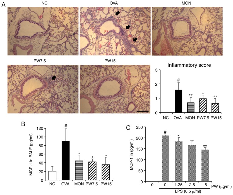
Figure 4.
Effect of PWRE on the influx of inflammatory cells into the lungs and on the downregulation of MCP-1 secretion in LPS-stimulated RAW264.7 macrophages. (A) Hematoxylin and eosin staining was used to determine the level of inflammatory cell influx (peribronchial lesion; magnification, ×100; scale bar, 50 µm) and the degree of the inflammation score was assessed by two independent observers. An ELISA was used to determine the MCP-1 secretion level in the (B) BALF samples of allergic asthma and in the (C) LPS-stimulated RAW264.7 macrophages. #P<0.05 vs. NC group; *P<0.05 and **P<0.01 vs. OVA group. PWRE, P. weinmannifolia root extract; MCP-1, monocyte chemoattractant protein-1; OVA, ovalbumin; MON, montelukast; BALF, bronchoalveolar lavage fluid; NC, negative control; LPS, lipopolysaccharide.

