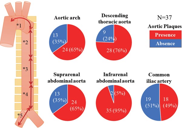
Fig. 2.

Distribution of all atherosclerotic plaques at each segment of the aorta
All atherosclerotic plaques were defined as yellow plaque, thrombus, or ruptured plaque.
Data are presented as n (%). The frequency of aortic plaques was the greatest at the IAA in the aorta (p < 0.001).
*1 (aortic arch); *2 (descending thoracic aorta); *3 (suprarenal abdominal aorta); *4 (infrarenal abdominal aorta); *5 (common iliac artery).
*1 vs. *4, p < 0.001; *2 vs. *4, p = 0.017; *3 vs. *4, p = 0.003; *4 vs. *5, p < 0.001.
