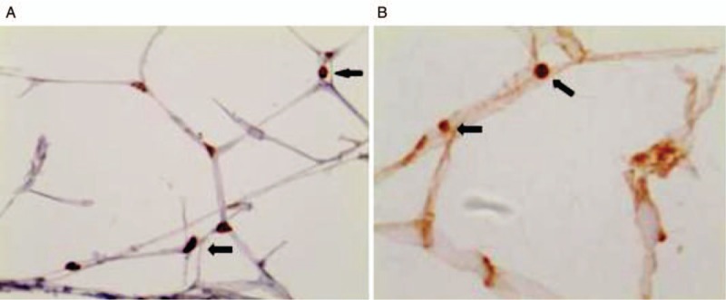
Figure 1.

Immunohistochemistry was used to observe the location of VDR and PPARγ in adipose tissue (Streptomyces antibiotic protein-peroxidase ligation staining, original magnification ×400). Brown-yellow granules (arrows) were seen in the adipose tissue nucleus, indicating the location of VDR (A) and PPARγ (B) in the adipose tissue nucleus. PPARγ: Peroxisome proliferator-activated receptor γ; VDR: Vitamin D receptor.
