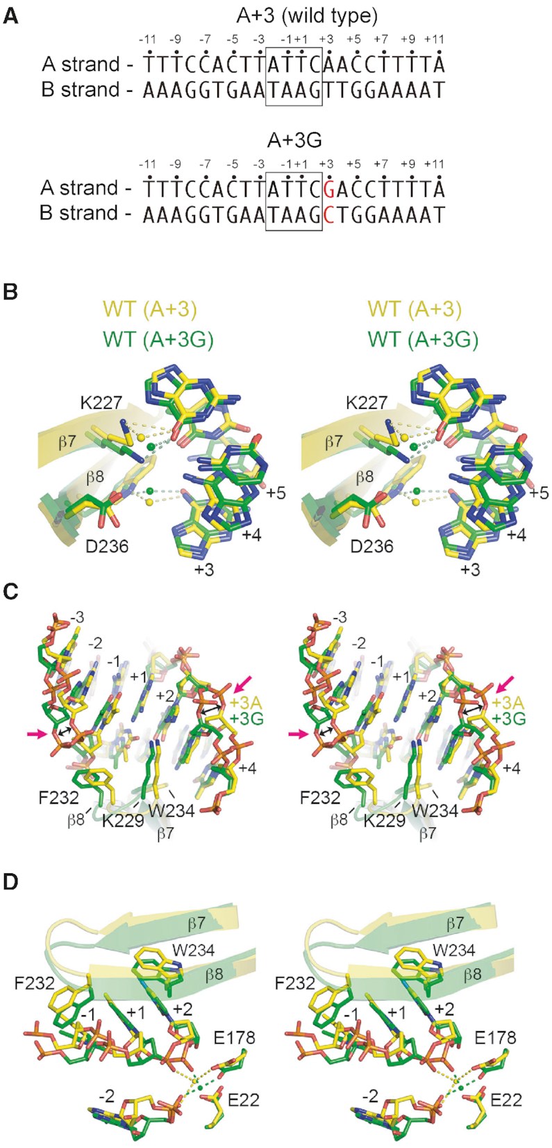
Figure 7.

Structures of wild-type I-OnuI with cognate A+3 and A+3G substrates. In all panels, the I-OnuI protein is colored according to substrate, with A+3 as yellow and A+3G as green. Note that the I-OnuI/A+G3 complex is post-cleavage and both DNA strands are cut. (A) Schematic of substrates used in crystallization experiment. (B) The network of direct and water-mediated contacts between K227, W234, D236 and bases +3, +4 and +5. (C) Distortions in the phosphate backbone of the A+3G substrate relative to the cognate substrate. (D) Perturbations of the active site residues E178 and E22 of the A+3G substrate relative to the cognate substrate. All panels are stereo images.
