
Figure 2.
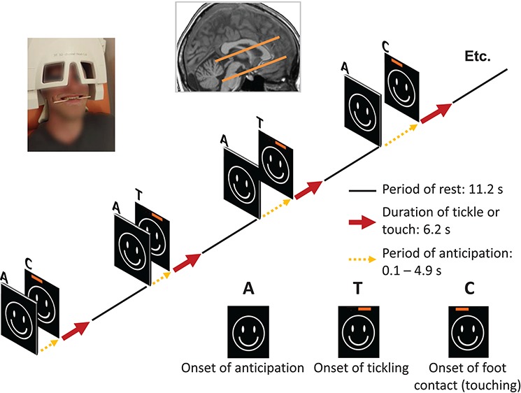
Experimental design. During the fMRI-scanning procedure, the participants experienced two different sensory stimulations, which were randomly applied: simple contact (C) or tickling (T) of the right foot. A visual cue signalized the upcoming stimulation, which followed only after a variable delay—the phase of anticipation (A)—of 0.1 to 4.9 s. To secure the unpredictability of the situation to the tickled person, the nature of the stimulation (tickling or touching) was signalized to the tickler alone by a superimposed red bar on the screen at the onset. The corresponding neuronal activity was measured in a defined brain volume, which included the AI, the hypothalamus and the PAG. A picture of a participant has been included, demonstrating the experimental set-up that was used.