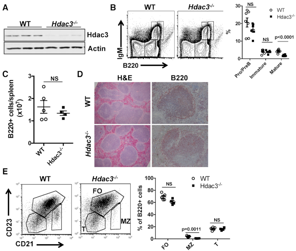
Figure 1.
B cell development is unaffected by CD19-cre driven deletion of Hdac3. (A) Loss of Hdac3 protein was determined by western blot analyses of proteins isolated from B220+ splenocytes of Hdac3+/+CD19cre+/−(WT) controls or Hdac3F/−CD19cre+/−(Hdac3−/−) animals. (B) Flow cytometric analysis utilizing IgM and B220 antibodies characterizes B cell development within the bone marrow of control and Hdac3-deleted mice. Graphical representation of B cell populations as a percentage of the total bone marrow from at least 6 mice per group is presented at the right. (C) Flow cytometry was used to determine the number of B220+ cells per spleen from multiple mice. (D) In order to characterize the structure of splenic follicles, formalin-fixed spleens were subject to H&E staining and immunohistochemistry with α-B220. (E) Flow cytometric characterization of splenic B cell populations was carried out. B220+ splenocytes were further analyzed based on surface CD23 and CD21 expression. Graph on right depicts five WT mice and four Hdac3−/− mice.

